
Urinary Stones
Urinary stones are crystal aggregates which form and grow in
urine. Urinary lithiasis is a very old disease: urinary
stones have been found in a 7,000 years old mummy.
The treatment of urinary stones is ancient as well. From the
"saxifraga" plant cooked in wine, used in the middle ages, to
the modern extracorporeal lithotripsy with shock waves,
mankind has been always troubled by urinary lithiasis.
Normally, several different substances (often crystals)

are present in urine, in a complex solution. The main
hypotheses on urinary stones formation are the
following.
Supersaturation.
An excess of concentration above a specific limit leads to
crystallization. The saturation threshold is dependent upon
temperature and acidity (pH) of the solution. An example of
this phenomenon can be observed adding salt to water. Beyond
a certain amount, the salt will not dissolve in water
anymore. When saturation limit of a substance in urine is
exceeded, crystal nucleation and growth will occur. After a
meal, an increased concentration of ossalate and calcium may
occur in urine. During the night, when fluid intake is low,
supersaturation may be reached. It is said that probably
urinary stones form at night.
Inhibitors: Even when high concentration and
supersaturation are reached, in some patients crystallization
doesn´t occur. This is due to some natural
substances which are present in urine, like some proteins,
magnesium and citrate. These substances may interfere with
crystallization. In certain patients there is a decreased
concentration of these inhibitors of crystallization. Citrate
and magnesium administration is used for urinary stones
prevention.
Crystal retention Generally crystals are
found in urine of healthy people, but only 10% of population
will form urinary stones. Anatomic and biochemical factors
can contribute to adhesion of small crystals to the urinary
mucosa.
Matrix: matrix is an organic amorphous
substance (a mucoprotein) which is present in urinary stones
in highly variable proportion. Though the role is uncertain,
some Authors have postulated that it can be involved in
urinary stones formation.
Urinary stones are usually made of several different layers,
that at section resemble the look of concentric growth of
trees.
Most stones (70-90%) contain calcium, together with oxalate,
phosphate, or carbonate. Struvite (magnesium ammonium
phosphate) are 15-20% of all stones, and often they are due
to urinary infection. Mostly they occur in women. Uric acid
stones (5-10%) are related to hyperuricemia; these stones are
radiolucent (they are not seen with plan x-rays) and often
they can be treated with medical therapy. Cystine stones are
rare (around 1%) and usually secondary to a congenital
metabolic defect. The incidence of urinary stones is
increasing in last decades. Bladder stones incidence,
prevalent in the past centuries, is now decreasing in western
countries, where an increment of upper urinary tract (kidney
and ureters) stones is observed.
The chance of being diagnosed with urinary stones along life
is 2.5-10%. About 20% of patients with urinary stones require
admission to a hospital. The age at first diagnosis is
between 20 and 50 years. Every patient with at least
one episode of urolithiasis should undergo periodic kidney
ultrasound, for the high risk of stone recurrence (50% at 10
years).
There is geographic and dietary variety in the incidence of
urinary lithiasis in different populations. Which are
risk factors for urinary stones?
*A diet rich with animal proteins and poor of vegetables and
fibers, like the ones in western countries
*A high intake of sodium
*Low fluid intake, especially in warm and dry climates (for
the risk of excess urinary concentration)
*Contrary to what people believe, a diet rich of calcium may
prevent urinary stones.
Several diseases can be associated to an increased risk of
stone formation, including some metabolic disorders, like
renal tubular acidosis, hyperuricemia, primary hyperoxaluria,
cystinuria, medullary sponge kidney. Hyperparathyroidism
(hyperfunction of parathyroid glands), sarcoidosis, some
tumors, especially of the blood (leukaemia, lymphoma), can
all increase the risk of urinary stones.
However, the majority of urinary stones (70%) are calcium
stones without apparent aetiology, with normal or increased
urinary calcium concentration.
About 15-20% of urinary lithiasis is related to urinary
infection, since the bacteria can alter the normal acidity of
urine.An increased risk of stone forming is associated to
immobilization, urinary tract obstruction, surgical urinary
diversion, bowel surgery for obesity, and to some drugs
therapy.
Symptoms and diagnosis
Urinary stones can cause different symptoms according to the
site. A kidney stone not obstructing the urine flow, can be
completely asymptomatic, or produce a mild flank pain.
The colic is due to a stone obstructing the passage of
urine, in the kidney or the ureters, with painful
overdistension cranial to the stone. In patients with a
stone, the risk of having colic is 40-50 %. The colic pain is
often very strong, intermittent, in the lumbar area or in the
flank. There can be nausea and vomit. When the stone is in
the ureters, the pain can irradiate toward the iliac region,
to reach the labia or the scrotum. When the stone is in the
distal ureter, it can mimic cystitis, with increased urinary
frequency and urgency.
Haematuria (blood in the urine) can be present in 40% of
cases. If there is a concomitant urinary infection, fever
with chills may occur. It is important to remember that
sometimes the urinary obstruction by the stone can be
partial, and the patient may be asymptomatic. It is mandatory
to undergo periodic ultrasound scan of the kidney, after a
first episode of urolithiasis.
Usually ultrasound scan of the bladder and kidney is the
first approach to urinary stones. Ultrasound scan is not
invasive, not painful, performed through a probe on the
abdomen. With the exam, the kidney

and the bladder can be visualized. Stones of the kidney, of the very first tract and last tract of the ureter can be diagnosed. The eventual distension (hydronephrosis)

of the kidney, due to obstruction of urine passage, can be
easily seen. However the whole ureter, which is a thin
tube passing behind the bowel, is hard to identify by
ultrasound.
Plan x-ray of the abdomen can show calcifications on the
kidney area, or along the probable way of the ureter. Since
more than 80% of stones contain calcium, they may be seen by
plan x-ray. Nevertheless, small or along the iliac bone
stones can be missed.
In these cases for decades urography has been the main test
for urinary stones diagnosis. It is performed through I.V.
injection of a contrast medium, containing iodine. Diagnosis
of iodine contrast adverse reaction and severe renal failure
are contraindications to urography. With urography all
excretory way can be seen on a 2 dimension film. Size and
site of the stones are diagnosed. Even radiolucent stones
(uric acid, struvite, cystine) can be seen as a filling
defect in the contrast.
In then last years urography has been gradually replaced by
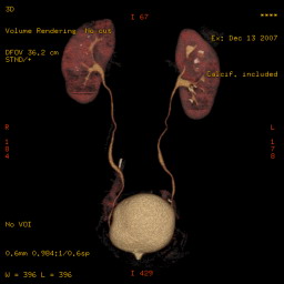
CT scan. Ct scan can be used without contrast medium,
providing accurate information about the size and the site of
the stone.When contrast medium is used, with last generation
CT scan 3D imaging of excretory ways can be obtained.
The aim of medical therapy is to prevent new stones formation
and the growth of already present stones. Only in selected
case medical therapy can dissolve stones. In patients with
pure acid uric stones, calcium free, (easily diagnosed
because they are radiolucent), urine alkalization can
dissolve even large stones. Urine is usually acid. Modifying
the pH (a number which is inversely proportional to the
acidity of a solution), acid uric stones can be dissolved in
3-6 months of therapy. The cheapest drug for urine
alkalization is sodium carbonate. It is contraindicated in
patients with hypertension (for increased intake of sodium).
Urine pH should be maintained at 6.5-7, for 3-6 months.
Allopurinol, a drug used to decrease uric acid in blood, can
be administered when hyperuricemia is present.
In patients with calcium stones, potassium citrate may
decrease the risk of stone recurrence. When hypercalciuria is
present, administration of thiazides (a group of diuretics)
can be effective for stone prevention. An increased fluid
intake is however essential in patients with urinary stones.
Even patients with large kidney stones can be asymptomatic,
until the stones obstruct the passage of urine.
Occasionally haematuria or a dull, mild flank pain may
occur.
When the first sign of a stone is a renal colic, the
treatment should be aimed to relieve acute pain. Non
steroidal anti-inflammatory drugs can be useful, while the
role of antispasmodic therapy is debatable.
When fever is present, antibiotic therapy should be started
(quinolones, or cephalosporines are indicated).
According to the site and size of the stone, often the stone
can be expelled spontaneously, with expectant approach, with
increased fluid intake and analgesic therapy when required
(tramadol drops can be a good option). The administration of
alpha-blockers can increase the chance of spontaneous passage
of the stone.
Frequently the colic is due to the passage of a stone along
the ureter, the thin tube connecting the kidney and the
bladder. Due to small calibre of the ureter, even a minute
stone can be obstructing.
The chance of spontaneous obstruction is highly depending on
the size of the stone.
< 4
mm
93%
4-6 mm
50%
7
mm
35%
>8
mm
0-5%
If a stone doesn´t pass within 2-3 weeks, the chance
of spontaneous expulsion drops considerably.
A ureter stone must be treated when:
- There is a severe hydronephrosis
- Doesn´t pass spontaneously within 2-3 weeks
- There is fever not responding to antibiotic therapy
- There is strong pain not responding to analgesics
- The size is such that spontaneous passing is very unlikely to happen
- In patients with single kidney or with bilateral ureteral stones
In case of hydronephrosis with fever and urinary infection,
urine should be drained from the kidney before stone
treatment. Urine can be drained through the excretory ways,
with a thin ureteral catheter, or with a tube through the
flank (percutaneous nephrostomy).
The ureteral catheter is introduced with a cystoscopy, with
a fiber optic, through the urethra. The catheter is pushed
upward until it reaches the kidney. Usually a double J stent
is used: it is called like this, because at both ends of the
catheter there are two pig-tails, to avoid displacement. The
ureteral catheter stays completely inside the body, between
the kidney and the bladder, thus by-passing the obstructing
stone.
Another option is the emergency drainage of urine through a
small tube placed inside the flank, directly into the kidney.
The tube (nephrostomy) is fixed to the skin by a stitch, and
the urine is collected in a bag. This is a temporary and fast
way to obtain drainage of infected urine.
Urinary stone treatment can be performed with several
techniques.
The least invasive approach to a stone is Extracorporeal
Shock Wave Lithotripsy (ESWL)
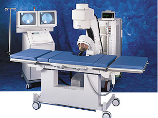
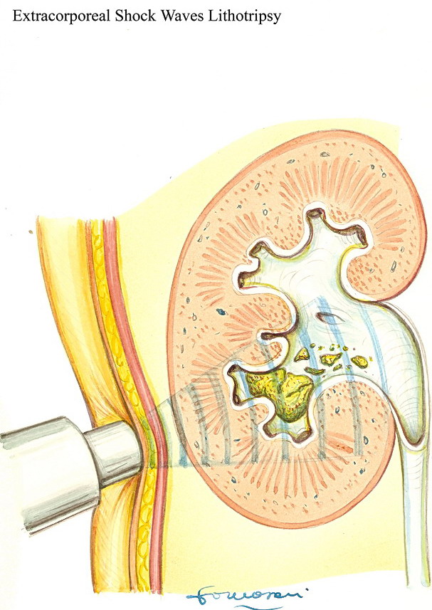
A special device produces intermittent shock waves, that are
transmitted to the body through a fluid medium (water, or a
gel patch). The waves are focused on the stone by ultrasound
and/or x-rays. According to size, site, and composition of
the stone, multiple procedures may be needed to obtain a good
fragmentation. The stone is not dissolved, but fragmented in
smaller stones, that can be expelled through the ureter for
their reduced size.
The limits of the procedure are related to the stone
structure (very hard stone can´t be fragmented), size
(stones larger than 20-25 mm are considered too large), or
site (stones near the iliac bone may not be treated properly)
For stones of the lower calyx of the kidney, success rate is
around 50-60%. For other kidney locations, the success is
higher, about 80-90%.
Though the risk of renal haematoma is low, it is higher for
patients with hypertension. Patients with severely impaired
coagulation shouldn´t undergo ESWL.
Some Authors have reported an increased risk of new
hypertension after ESWL, but the data are contradicting.
Recently it has been suggested that ESWL might facilitate the
onset of diabetes.
When kidney stones are too large for ESWL treatment, the
technique of choice is percutaneous lithotripsy. With the
patient in prone position, the excretory ways of the kidney
are accessed with a needle through the flank. A fiber optic
instrument is introduced and the stone is reached.
The stone is fragmented under vision, with ultrasound,
laser, or mechanic energy. Major fragments are removed
through the instrument. The operation requires general
anaesthesia. Success rate is above 90%. Complications are
rare, but they may involve renal bleeding.
Stones of the ureter can be treated with ESWL or with
ureteroscopy (endoscopy). A thin fiberoptic instrument, about
3 mm in diameter, is introduced through the urethra, the
bladder, and up into the ureter, until the stone is met. With
the same forms of energy available for percutaneous
lithotripsy, the stone is fragmented. The patient can be
dismissed in the next 24 hours. The procedure is successful
in 85-95% of cases.Open surgery is today very rare, and it is
reserved to 2-3% of urinary stones. Usually patients with
staghorn stones, filling all the kidney excretory ways, are
candidates. In patients with large ureter stones, not
responding to ESWL , after failure of ureteroscopy, a
laparoscopic approach can be used.
Bladder stones are rarer, and usually they are related to
chronic urinary retention, due to voiding obstruction, like
in benign prostatic hyperplasia or in urethral strictures.
Urinary infection and indwelling catheters are risk factors
for bladder stones. Bladder stones can be treated with
endoscopic approach, with a fiberoptic instrument through the
urethra. Mechanical fragmentation is performed. Very large
stones may require surgical approach for removal.